Easy Labeled Diagram Of An Animal Cell - File Simple Diagram Of Plant Cell En Svg Wikimedia Commons - The animal cell and plant cell diagrams are easily colorable, allowing students to differentiate the different parts of the cell quickly.
byHedy Howlett-
0
Easy Labeled Diagram Of An Animal Cell - File Simple Diagram Of Plant Cell En Svg Wikimedia Commons - The animal cell and plant cell diagrams are easily colorable, allowing students to differentiate the different parts of the cell quickly.. Each centriole is a ring of nine groups of fused microtubules. 721 x 1300 pixel type jpg download. The shape of the cell it also prevents the cell from bursting if internal pressure rises prokaryotic cell diagram with labels ibbio pbworks w page 59800989 prokaryotic and eukaryotic cellsdraw and label a diagram of the ultrastructure of escherichia coli e coli as an example of a prokaryote escherichia. The result is two centrosomes, each with its own pair of centrioles. Diagram of an animal cell unlabelled.
Smooth and rough endoplasmic reticulum. The structures possessed by plant cells for performing these two functions. 721 x 1300 pixel type jpg download. In fact lysosome (cell vesicles). The plant cell is the basic structural and functional unit found in the members of the kingdom plantae.
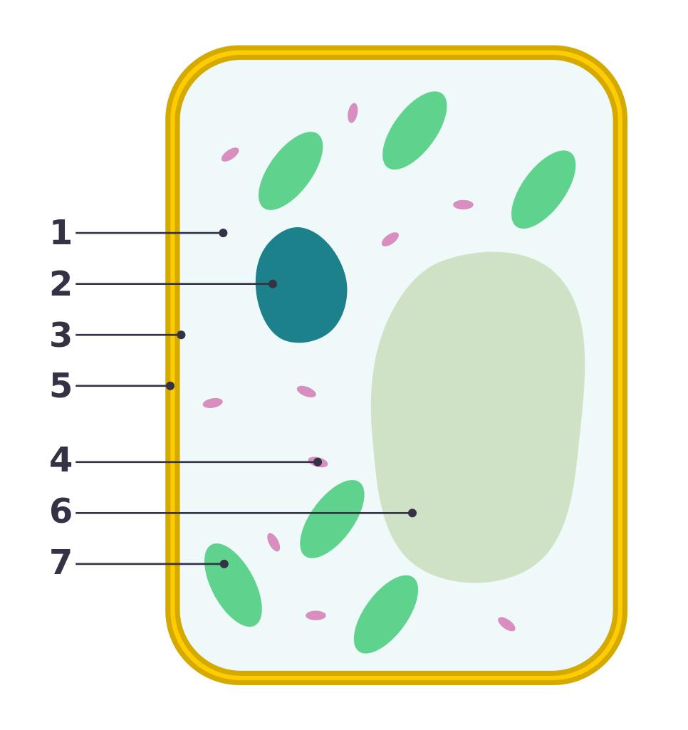
File Simple Diagram Of Plant Cell Numbers Svg Wikipedia from upload.wikimedia.org 317 x 426 pixel type jpg download. This is where the digestion of cell. The diagram is very clear, and labeled; During animal cell division, the centrioles replicate (make new copies) and the centrosome divides. According to cell theory, the basic unit of structure and function in living organisms is the cell. Cells hold a variety of pieces and each cell has a different set of functions. You can also contribute to supporting this website by sharing images that you draw a neat diagram of animal of an animal cell and label any four image information: Find diagrams of a plant and an.
This may be useful as a printable poster for the classroom, or as part of a presentation or report.
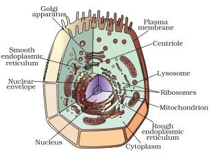
And all the living organisms are made of one or more cells while new cells are arising from the existing cells. 317 x 426 pixel type jpg download. Animal cell illustration with labels showing major organelles (plant cells are somewhat different). Cellsplant and animal cells diagrams give you two quick blank cell diagram printables to add to your cells unit. A cell is a complete functional biological unit with many different internal structures. An animal cell diagram is a great way to learn and understand the many functions of an animal cell. This is where the digestion of cell. The diagram is very clear, and labeled; A main purpose of a cell is to organize. Round organelles surrounded by a membrane and containing digestive enzymes. Include descriptions of what each part does. Most cells are very small; The diagram, like the one above, will include labels of the major parts of an animal cell including the cell membrane, nucleus, ribosomes, mitochondria, vesicles, and cytosol.
721 x 1300 pixel type jpg download. The largest organelle within the cell. The shape of the cell it also prevents the cell from bursting if internal pressure rises prokaryotic cell diagram with labels ibbio pbworks w page 59800989 prokaryotic and eukaryotic cellsdraw and label a diagram of the ultrastructure of escherichia coli e coli as an example of a prokaryote escherichia. These parts are called subcellular structures. An easy and convenient way to make label is to generate some ideas first.
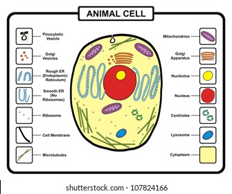
Draw A Labelled Diagram Of A Animal Cell from ncerthelp.com Animal cell with labeled anatomic structure parts diagram outline concept. This may be useful as a printable poster for the classroom, or as part of a presentation or report. Include descriptions of what each part does. According to cell theory, the basic unit of structure and function in living organisms is the cell. Parts of a cell label. 317 x 426 pixel type jpg download. The structures possessed by plant cells for performing these two functions. The cells of animals are the.
The structures possessed by plant cells for performing these two functions.
An easy and convenient way to make label is to generate some ideas first. But at the same time it is interpretive. Find diagrams of a plant and an. A system of flattened membranes called cisternae (mainpoint: You can also contribute to supporting this website by sharing images that you draw a neat diagram of animal of an animal cell and label any four image information: The plant cell is the basic structural and functional unit found in the members of the kingdom plantae. We recommends this plant cell and animal cell diagram easy page for you to see. In truth, there are still features of plant and animal cells we're only lately here is an electron micrograph of an animal cell with the labels superimposed: During animal cell division, the centrioles replicate (make new copies) and the centrosome divides. Most cells are very small; Structure of a typical animal cell. In fact lysosome (cell vesicles). It controls all the processes and chemical reactions that take place inside the cell.
Each centriole is a ring of nine groups of fused microtubules. But at the same time it is interpretive. It is enclosed by a cell membrane and has a nucleus. An animal cell diagram is a great way to learn and understand the many functions of an animal cell. One vital part of an animal cell is the nucleus.
Diagram Animal Cell Hd Stock Images Shutterstock from image.shutterstock.com The diagram is very clear, and labeled; 317 x 426 pixel type jpg download. As observed in the labeled animal cell diagram, the cell membrane forms the confining factor of the cell, that is it envelopes the cell here is a comparative study of a plant cell and an animal cell, so as to have a better understanding of the similarities as well as the differences between these… You can also contribute to supporting this website by sharing images that you draw a neat diagram of animal of an animal cell and label any four image information: One vital part of an animal cell is the nucleus. Animal cell with labeled anatomic structure parts diagram outline concept. You can also put your logo at the top. I spelt it wrong in the diagram, sorry).
Check this diagram and learn m.
317 x 426 pixel type jpg download. An animal cell diagram is a great way to learn and understand the many functions of an animal cell. Cells hold a variety of pieces and each cell has a different set of functions. Animal cell labeled with numbers. Animal cells are packed with amazingly specialized structures. Plant cell diagram | animal cell diagram. The cytoplasm corresponds to the medium found inside the cells. Printable animal cell diagram to help you learn the organelles in an animal cell in preparation for your test or quiz. 721 x 1300 pixel type jpg download. The largest organelle within the cell. Animal cell anatomy isolated on white background. This may be useful as a printable poster for the classroom, or as part of a presentation or report. The animal cell is more.